Northwestern Medicine Palos Hospital Now Offering Cardiac MRI

![]()
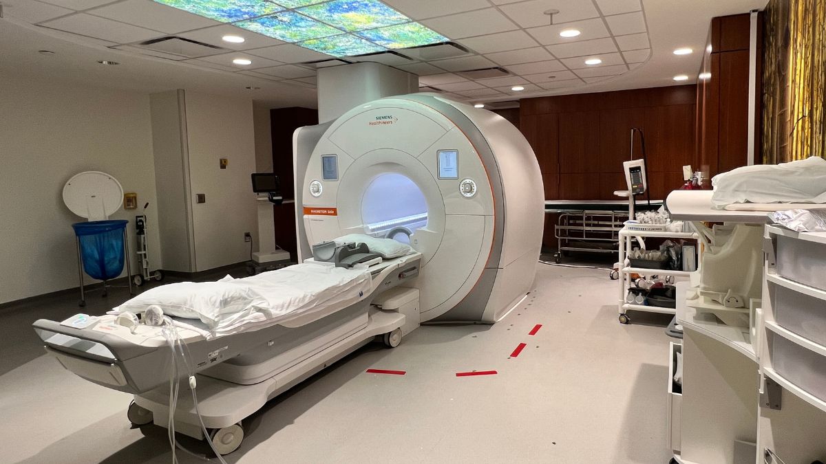
Northwestern Medicine Palos Hospital Now Offering Cardiac MRI (Palos Heights, IL) — Cardiac MRI is now available at Northwestern Medicine Palos Hospital. The versatile tool is used to diagnose many cardiac conditions and is the best test available to understand in full detail the structure of the heart.
Northwestern Medicine’s imaging systems are fully integrated across the health system’s campuses. This allows radiologists practicing at Northwestern Medicine Palos Hospital to easily collaborate electronically with colleagues across the health network, including downtown at Northwestern Memorial Hospital.
“World-class researchers in cardiac MRI at Northwestern Medicine work hard every day to advance the field so that patients can benefit from more accurate diagnosis, which leads to better treatments,” said R. Kannan Mutharasan, MD, medical director of the Northwestern Medicine Bluhm Cardiovascular Institute at Palos Hospital.
Dr. Mutharasan and Bradley Allen, MD, MS, chief of Cardiovascular and Thoracic Imaging at Northwestern Medicine, answer some common questions about cardiac MRI.
Why would a cardiologist order a cardiac MRI?
Cardiac MRI can test for multiple indications including evaluation of heart failure, potential cardiac masses, valve disease, a blood clot in the heart, or acute cardiac injury such as myocarditis. It can also be used as part of an arrythmia work-up or treatment planning prior to ablations.
What information can a cardiac MRI reveal that other testing may not?
“Cardiac MRI is considered the gold-standard for evaluation of heart size and function. In addition, MRI is excellent at tissue characterization and can provide information about scar or inflammation to the heart muscle,” said Dr. Allen.
More advanced types of MRI like stress perfusion can help determine if there is reduced blood flow to the heart, while a special type of MRI called phase contrast imaging can visualize and measure blood flow and velocity. This information can diagnose various conditions like heart attack, myocarditis, or heart valve disease; while also helping to estimate risk and potential success of various treatment strategies.
“Hypertrophic cardiomyopathy, a genetic condition that affects up to 1 in 500 individuals, is a good example of a condition that a cardiac MRI may pick up, but a more routine cardiac test like an echocardiogram may miss,” said Dr. Mutharasan. “Understanding whether someone has hypertrophic cardiomyopathy is important because not only does it affect the individual patient, but it may impact the health of family members who may have the condition but not realize it.”
How does the test work? How long does it take?
MRI generates images using a very strong magnetic field. Patients lie flat and are placed in the center of the MRI scanner which looks like a round tube. A special piece of equipment called a coil is placed on their chest.
“The MRI machine makes many loud sounds and vibrations which is the machine producing small magnetic field changes within the patients. The coil on the chest functions like an antenna which receives the magnetic signals coming from the patient,” said Dr. Allen.
These signals are sent to a computer where they are used to build an image of the part of the body being scanned. The images are then reviewed by a cardiac imaging expert. Most cardiac MRIs require intravenous injection of gadolinium contrast agent. The test takes 60-90 minutes to complete.
Are there any risks?
Because the MRI machine uses very strong magnets, patients cannot bring anything metal into the scanner. All patients will be screened for metal implants or other metal objects that could be unsafe in the scanner. Patients with ICD or pacemakers usually can undergo MRI but require special settings to be used on their device. There is no ionizing radiation associated with MRI so pregnant women can undergo MRI safely but cannot receive gadolinium. Patients with poor kidney function may not be eligible to receive gadolinium.
What is new in the world of cardiac MRI?
“Cardiac MRI is a very exciting research space. Some work is focused on how to make the imaging faster so the studies can be completed in 30 minutes or less,” said Dr. Allen. “Additional work has focused on how our ability to characterize changes in the heart tissue can help better define patient diagnoses and risk in diseases like cardiac amyloidosis and in patients with heart transplant.”
Evaluating cardiac blood flow is another area of expertise at Northwestern Medicine using a tool called 4D flow MRI. Machine learning and artificial intelligence is starting to significantly impact cardiac MRI in a whole range of applications including imaging the patient, performing analysis and quantification, and generating reports.
When should I ask my doctor about cardiac MRI?
The role of Cardiac MRI should be discussed with your cardiologist and is especially indicated if the underlying cardiac disease or extent of disease is not clear after evaluation with other diagnostic tools such as echocardiography.
To make a cardiology appointment at Northwestern Medicine Palos Hospital, please call 708-923-4200 or visit heart.nm.org.
Northwestern Medicine Palos Hospital Now Offering Cardiac MRI


Responses